
Methanogens
Predicting the Function of Possible Radical S-adenosyl-L-methionine Enzymes in Methanocaldococcus jannaschii using Bioinformatic and Computational Methods
Abstract
It is believed that radical S-adenosyl-L-methionine (SAM) enzymes (a special class of iron-sulfur binding clustered proteins) played a crucial role in the first metabolic pathways to evolve on this Earth. Very little is known about this class of proteins, but we hypothesize they play a crucial role in methanogenic sulfur metabolism, a biochemical pathway currently unknown to science. Some of these reactions are likely carried out by members of the duf39 gene family, which includes MJ0100 and MJ1681. These genes as well as MJ0542 and their homologs were computationally analyzed to better predict their functions. The investigation included sequence alignment, structure modeling, energy minimization, and energy scoring, with the goal of finding conserved cysteines. These conserved cysteines give us insight into the mechanisms of these proteins. We hope to shed light on this metabolic pathway through these computational methods.
Introduction
Radical SAM enzymes were first discovered in 2001 and are defined as a protein superfamily with over 600 members—since then, this superfamily has grown tremendously. These enzymes’ mechanisms involve an iron-sulfur cluster bound to precisely three cysteines (any more and the protein is likely a ferredoxin, a family of iron-sulfur cluster proteins with four cysteine motifs). Radical SAM enzymes carry out many different functions, including methylations, sulfur insertions, and protein radical formation, yet many functions of this unusual superfamily remain unknown.
The first motif discovered was CX3CX2C, found in over 90% of the originally researched proteins. The remainder had variances of that motif, which has since expanded to include CX2CX4C, CX5CX2C, CX14CX2C, CX2CX2FC, CX2CX2YC, and CX2CX27C. The three-cysteine motif works by binding an iron-sulfur cluster at its center, allowing for the transfer of an electron.
Methanocaldococcus jannaschii was discovered by a submarine at a hydrothermal vent and was the first methanogen to have its genome sequenced, in 1996. M. jannaschii has genes connected to all three domains of life. We know that, to produce energy, M. jannaschii reduces CO2 into CH4 (methanogenesis). But we assume that, for survival, M. jannaschii also oxidizes sulfur in order to reduce toxicity and produce essential compounds such as 4Fe-4S binding clusters and Coenzyme M. Unfortunately, these pathways are currently unknown; we hypothesize the duf39 gene family is involved, and we have experimentally proven MJ0542 is able to metabolize hydrogen sulfide — but the question is, how?
duf39 Gene Family: MJ0100 and MJ1681
The duf39 gene family encompasses over 1200 proteins, including MJ0100 and MJ1681. Like M. jannaschii, this gene family is usually expressed as two distinct proteins in methanogens. It is known through sequence analysis that one contains two ferredoxin motifs and the other possesses a CBS domain, but the functions of these proteins remain hidden. We hypothesize they may be involved in the steps between hydrogen sulfide’s first metabolism and the production of a sulfone group.
Sulfur Metabolism: MJ0542
Because hydrogen sulfide readily diffuses across a cell membrane and can be toxic, we figured there must be some pathway utilizing this massive amount of sulfur. Methanogens have somehow been metabolizing this sulfur into organic compounds, iron-sulfur binding clusters, and maybe even elemental sulfur. Experiments have shown the enzymes responsible for this metabolism are MJ0542 (PEP synthase), MJ1453, and their Methanocaldococcus fervens homologs. We hope to use sequence alignment and structure modeling to better predict the unknown functions of MJ0542.
Methods
These methods pertain to MJ0100, MJ1681, and MJ0542, referred to below as “the proteins.”
BLAST / UniProt
BlastP searched the Archaea, Bacteria, and Eukaryota databases separately (E-threshold 10, BLOSUM-62, no filtering, gaps allowed). The top 25 results per domain were aligned, and every cysteine was tallied by hand to score conservation.
I-TASSER & SWISS-MODEL
I-TASSER threaded each sequence against the PDB and clustered candidate folds (SPICKER) to return five models per protein, along with the top ten structurally similar PDB files and top five probable ligands. Each model was validated with SWISS-MODEL (ANOLEA, QMEAN/QMEAN6, DFire, PROCHECK, DSSP, Promotif), energy-minimized in Maestro, and validated again.
PyMOL
Cysteine locations and inter-sulfur distances were measured on the minimized models to look for plausible 4Fe-4S binding sites. All protein renders on this page came from PyMOL.
SIFTER
Statistical Inference of Function Through Evolutionary Relationships predicts function from phylogenetic placement among homologs of known function, returning a confidence score from 0 to 1.
phyloT / UniProt
phyloT built a taxonomy-based phylogenetic tree of every duf39 gene in methanogens (27 UniProt IDs couldn’t be mapped to an NCBI ID). A second tree of MJ0542 homologs was built later in UniProt from the top 25 BLAST hits per domain.
Results
Sequence-based homolog search and alignment
No possible function could be predicted from the BLAST results for MJ0100. Sequence alignment showed no cysteine conservation with the eukaryote results. Two cysteines were fully conserved between the archaea and bacteria results (C54 and C133), and two more were partially conserved among the archaea (C159 and C287).
Structure modeling and model validation
Energy minimization and model validation determined model 1 was most favorable. Toggle between the SWISS-MODEL validation plots below to see the effect of Maestro energy minimization on local quality scores.
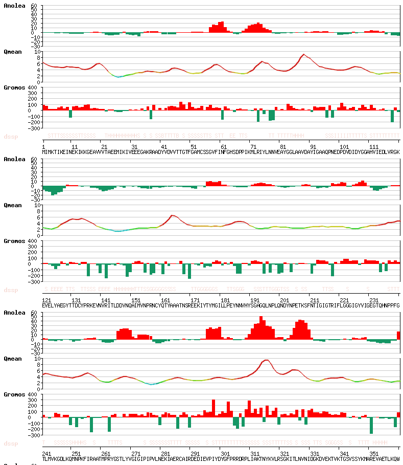
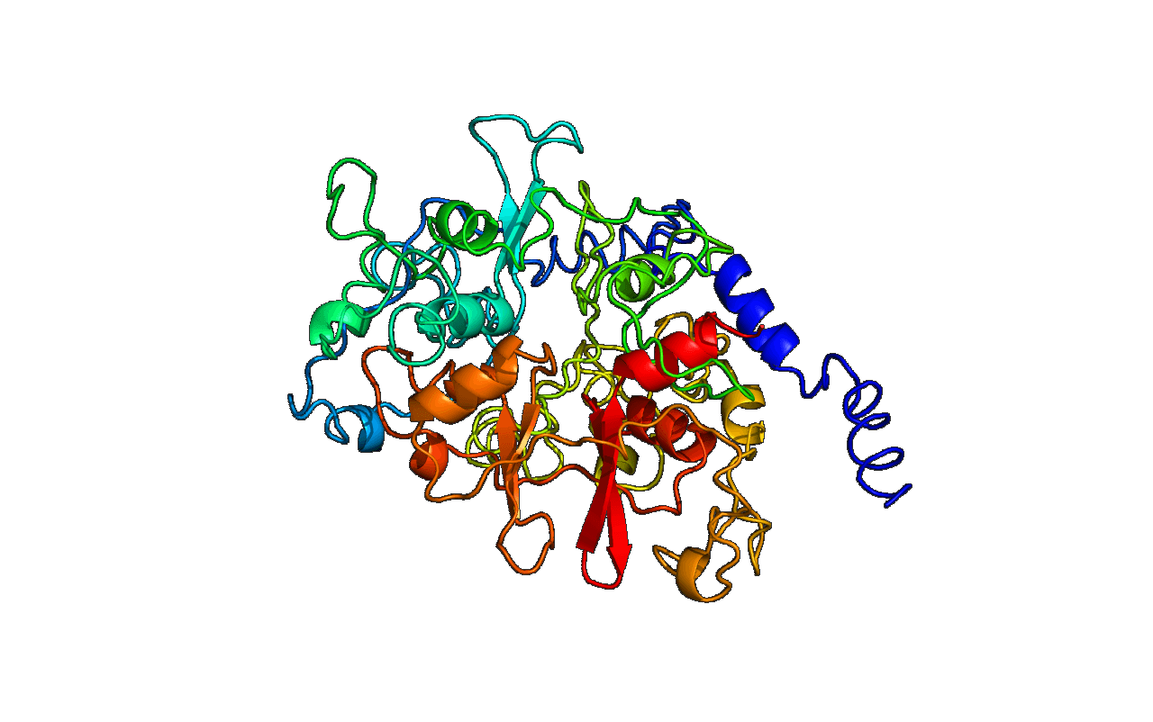
The top five of I-TASSER’s top ten homologous PDB structures suggest MJ0100 may be an AMP-binding protein — the top four are specifically AMP-activated kinases, and the top three predicted ligands are adenosines with varying numbers of phosphates. No cysteines are close enough to bind an iron-sulfur cluster.
Function prediction
SIFTER returned 39 possible functions with a confidence score of just 0.01, but also predicted cystathionine beta-synthase (CBS) activity with a confidence score of 0.94. CBS domains catalyze the production of L-cystathionine from L-serine and L-homocysteine, a crucial step in cysteine metabolism, and are known to be regulated by SAM. MJ0100’s CBS domain is likely used to regulate the other domain’s activity rather than catalyze a reaction itself.
Phylogenetic Trees
MJ0542 homologs
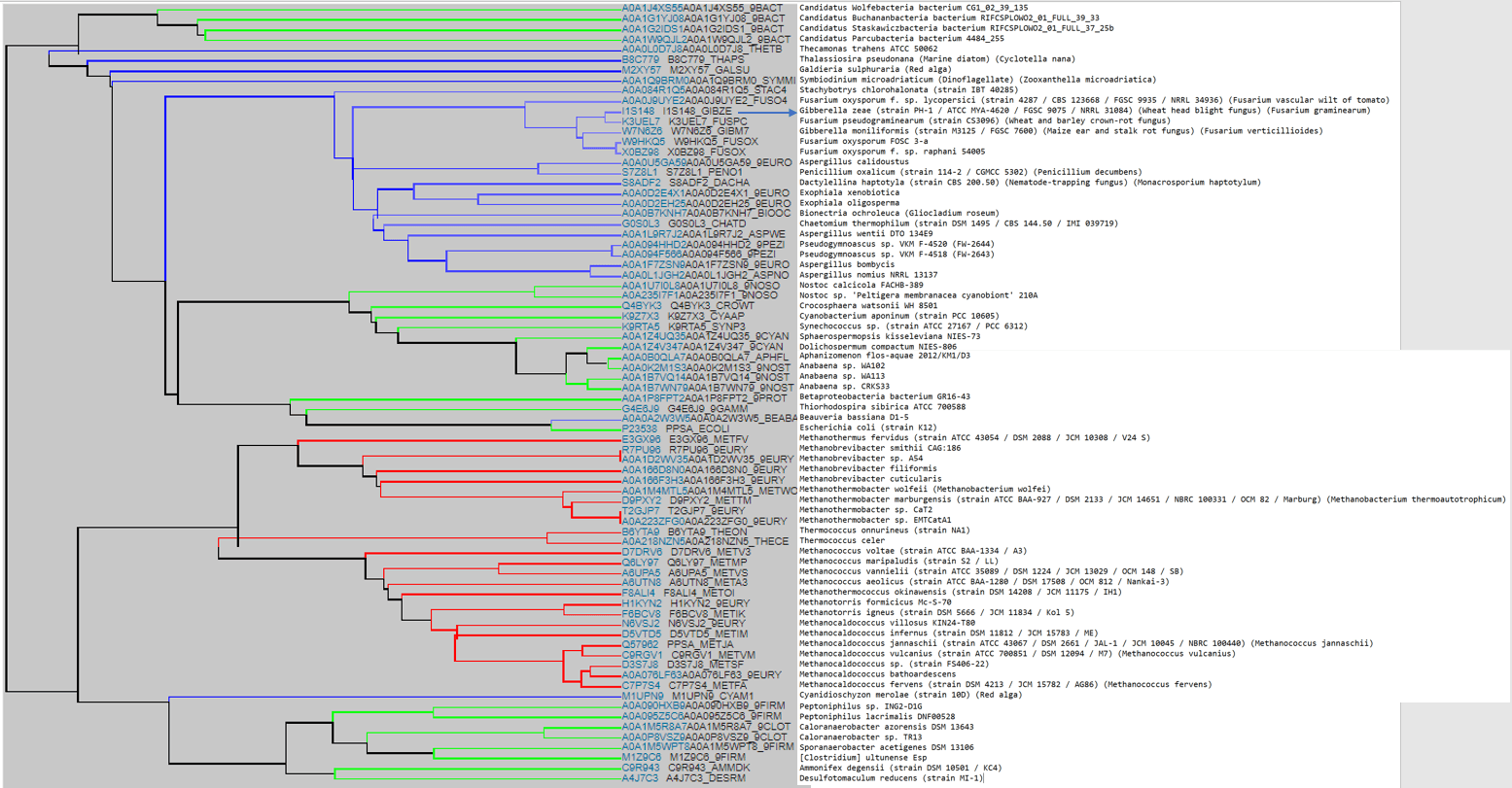
This tree was refined to exclude repeated species and to include E. coli’s closest homolog to MJ0542 — toggle between the original and revised version below.
Discussion and Future Work
MJ0100
Based on the PDB analogs given by I-TASSER, it’s possible MJ0100 is involved in adding a sulfone group to some organic molecule — plausible if MJ0100 actually binds an adenosine 5-monosulfate instead of an adenosine 5-monophosphate like its RCSB analogs. Based on experimental evidence, it’s more likely the CBS domain regulates MJ0100 instead of doing any reactions itself.
MJ1681
Much more research is needed. The iron-sulfur binding sites seem to be in an exposed position, which would weaken any binding affinity. The two motifs also seem to play off each other, possibly pointing to a new extended motif yet to be described. The model could also simply be inaccurate, despite favorable energetics.
MJ0542
Based on the SIFTER results, it’s possible MJ0542 is a dimer, which would make it harder to determine its hidden function(s). Sequence alignment highlighted the most essential cysteines, but visualizing their positions in PyMOL didn’t produce a breakthrough. The next step is molecular docking.
Phylogenetic Trees
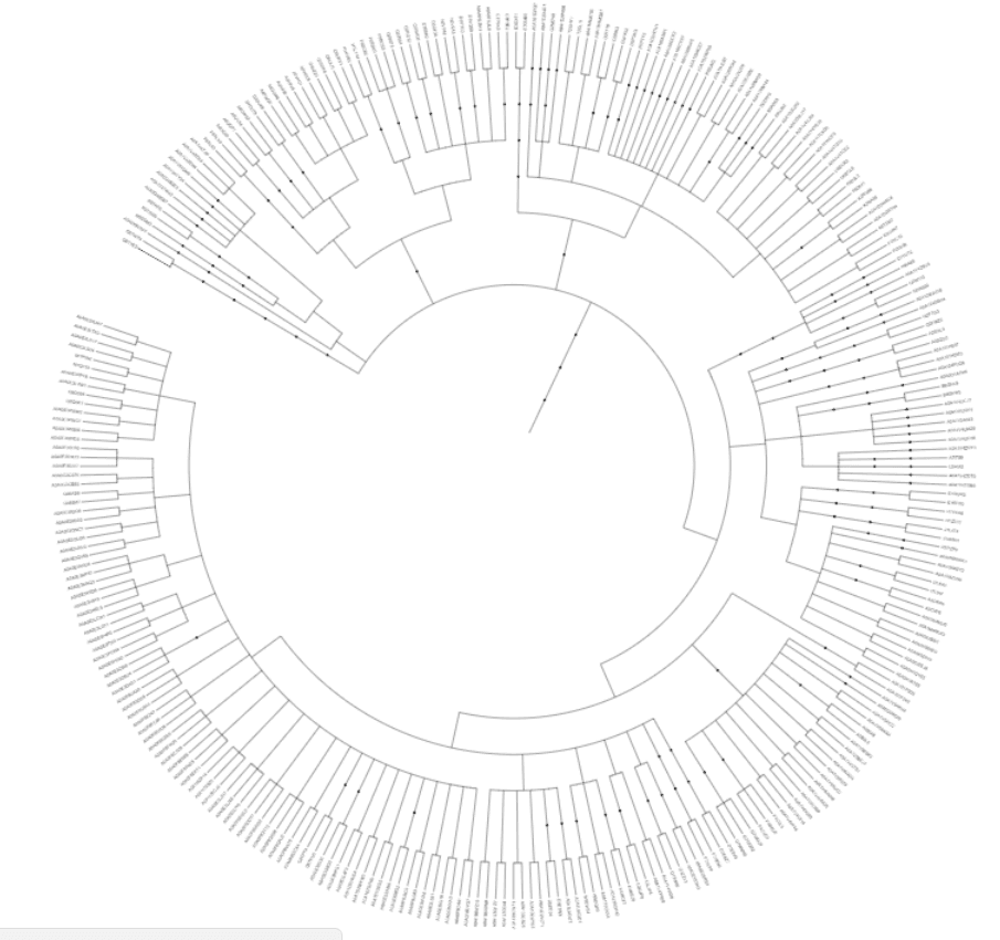
The duf39 tree shows how these two genes are shared among methanogens; adding sequence lengths to it could show that when an organism has two duf39 genes, it’s most likely to have one sequence below 450 amino acids paired with one above 450. MJ0542’s tree made the clades easier to follow, but UniProt seemed to mis-organize some bacteria and eukaryotes — the next step is rebuilding it in phyloT with more diverse organisms.
More research is needed to determine these proteins’ functions with confidence. Molecular docking and identifying key binding-site residues will be essential next steps.
References
- Sofia, H. J., Chen, G., Hetzler, B. G., Reyes-Spindola, J. F., and Miller, N. E. (2001) Radical SAM, a novel protein superfamily linking unresolved steps in familiar biosynthetic pathways with radical mechanisms, Nucleic Acids Res. 29, 1097–1106.
- Jones, W., Leigh, J., Mayer, F., Woese, C., and Wolfe, R. (1983) Methanococcus jannaschii sp. nov., an extremely thermophilic methanogen from a submarine hydrothermal vent, Arch. Microbiol. 136, 254–261.
- Reeve, J. N., Nölling, J., Morgan, R. M., and Smith, D. R. (1997) Methanogenesis: genes, genomes, and who’s on first?, J. Bacteriol. 179, 5975.
- White, D. M. R. H. (2017) Sulfide metabolism in Methanocaldococcus jannaschii: Promiscuity of a phosphoenolpyruvate synthase? (Tech, V., Ed.).
- (2018) UniProt, UniProt Consortium.
- Mathai, J. C., Missner, A., Kügler, P., Saparov, S. M., Zeidel, M. L., Lee, J. K., and Pohl, P. (2009) No facilitator required for membrane transport of hydrogen sulfide, PNAS.
- Roy, A., Yang, J., and Zhang, Y. (2012) COFACTOR: an accurate comparative algorithm for structure-based protein function annotation, Nucleic Acids Res. 40, W471–W477.
- Yang, J., and Zhang, Y. (2015) I-TASSER server: new development for protein structure and function predictions, Nucleic Acids Res. 43, W174–W181.
- Zhang, Y. (2009) I-TASSER: Fully automated protein structure prediction in CASP8, Proteins: Structure, Function, and Bioinformatics 77, 100–113.
- Benkert, P., Tosatto, S. C., and Schwede, T. (2009) QMEANclust: estimation of protein model quality by combining a composite scoring function with structural density information, BMC Structural Biology 9, 35.
- Schrödinger, L. (2010) PyMOL The PyMOL Molecular Graphics System, Version.
- (2018) SIFTER, UC Berkeley.
- (2018) phyloT, Ivica Letunic.